

Echocardiography is an imaging method that uses sound waves to help evaluate the structure of the heart and how it functions. This method provides detailed information about the size of the heart chambers, the movement of the heart walls, pumping strength, the structure and function of the heart valves, and blood flow.
Echocardiography can be used to evaluate heart failure, heart valve diseases, congenital heart problems, and certain conditions affecting the heart muscle. The images obtained can provide the physician with useful information during diagnosis and follow-up. This method may also be used, when necessary, to monitor the response to treatment.
Where is Echocardiography Performed?
Echocardiography is an imaging method that uses ultrasound waves to evaluate the structure and function of the heart. The answer to the question “Where is echocardiography performed?” may vary depending on the type of echocardiography required and the patient’s needs. Echocardiography is generally performed in hospital cardiology departments, medical centers, and healthcare facilities that provide medical imaging services.
Standard transthoracic echocardiography (TTE) is performed through the chest. During the procedure, gel is applied to the chest and an ultrasound probe is placed at different points to examine the heart chambers, heart valves, wall movements, and blood flow. The images obtained during the procedure can provide information about the structural and functional characteristics of the heart.
Echocardiography may be performed in the following healthcare settings:
- Hospital cardiology departments
- Cardiology clinics and medical centers
- Medical imaging centers
- Healthcare facilities offering echocardiography services
Denge Tıp is one of the medical imaging centers offering echocardiography services. The procedure can be planned based on the patient’s symptoms and medical assessment. The procedure can be planned according to the patient’s symptoms and medical evaluation. At Denge Tıp, echocardiography can be used to assess the structure and functional characteristics of the heart.
In some cases, different echocardiography methods, such as transesophageal echocardiography (TEE), may be required. Since this method obtains images through the esophagus, it should be performed at a healthcare facility with the appropriate equipment and medical facilities.
Where echocardiography is performed is important, but determining which type of echocardiography is appropriate is equally important. Therefore, the patient’s symptoms and clinical condition are evaluated before the procedure to determine the most suitable method.
How is Echocardiography Performed?
Echocardiography is most commonly performed using the transthoracic method through the chest. Electrodes may be placed on the chest to monitor the heart rhythm simultaneously with the images. Gel is then applied to the chest to facilitate the transmission of ultrasound waves. Images of the heart are obtained from different angles using an ultrasound probe called a transducer.
The main steps followed during the procedure are as follows:
- The probe is placed on different areas of the chest to obtain images from various sections of the heart.
- The person may be asked to turn onto their left side to obtain more suitable images.
- During some parts of the examination, the person may need to change their breathing pattern or briefly hold their breath.
- Doppler echocardiography may be used to evaluate the direction and speed of blood flow within the heart.
- The images and measurements obtained are evaluated by the physician after the examination.
Standard transthoracic echocardiography does not use ionizing radiation and, in most cases, does not require special preparation. The basic steps followed during echocardiography are similar, but the details of the procedure may vary depending on the person’s age, health status, and level of cooperation during the examination. The process may also differ in certain respects for infants, children, and adults.

In Infants
In infants, echocardiography is most commonly performed using the transthoracic method, meaning through the chest. First, the infant is placed in an appropriate position. Gel is then applied to the chest, and a small ultrasound probe is moved over different areas to obtain images of the heart. When necessary, the probe may also be used near the abdominal area to obtain additional images.
For clear images to be obtained, it is important for the infant to remain as calm and still as possible during the examination. During the procedure, healthcare professionals may use various methods to help the infant relax. If the infant’s movement makes imaging significantly difficult, the physician may assess whether sedation is needed in some cases. Standard transthoracic examination uses sound waves and does not involve radiation.
In Adults
During echocardiography, an adult usually lies on an examination table, and most images are obtained while the patient is positioned on their left side. After gel is applied to the chest, the ultrasound probe is placed at specific points to visualize different areas of the heart. Throughout the examination, the position and angle of the probe are adjusted, and different views are recorded.
While some images are being obtained, the person may be asked to take a deep breath, briefly hold their breath, or change position. The Doppler method may also be used to examine blood flow through the heart valves and chambers. Standard transthoracic echocardiography generally does not require special preparation. After the examination is completed, the recorded images and measurements are evaluated together by the physician.
In Children
In children, echocardiography is essentially performed using the same transthoracic method used in adults. During the procedure, the child lies on an examination table, gel is applied to the chest, and the ultrasound probe is moved over different areas to obtain images of the heart from various angles. The child is asked to remain as still as possible during the procedure, which is important for obtaining clearer and more accurate images.
How Long Does Echocardiography Take?
The duration of echocardiography may vary depending on the method used and the scope of the examination. Standard transthoracic echocardiography is generally completed in approximately 30–60 minutes. During this time, images of the heart are obtained from different angles and the necessary measurements are taken. When needed, the Doppler method may also be used to evaluate blood flow within the heart.
The main factors that may affect the duration of echocardiography include:
- The need to obtain detailed images from different areas of the heart,
- The need for additional measurements or Doppler assessment,
- The need to repeat images from different angles,
- The person changing position during the procedure,
- Infants or children having difficulty remaining still during the procedure,
- The use of a type of echocardiography other than the standard transthoracic method.
A longer procedure does not, by itself, mean that there is a problem with the heart. In some cases, images may need to be repeated to achieve adequate image quality or to examine certain structures in greater detail. The need to evaluate the heart valves, heart chambers, or heart walls more closely during the examination may also increase the total duration.
In addition, with different methods such as stress echocardiography and transesophageal echocardiography, the total duration may vary depending on the preparation, procedure, and follow-up stages. Determining the method to be used before the procedure can help provide the person with a clearer estimate of the approximate duration.
What Findings Can Be Obtained?
After echocardiography, various findings related to the structure and function of the heart can be obtained. During the examination, the overall appearance of the heart, as well as the size of the chambers, movement of the heart muscle, function of the valves, and blood flow within the heart are evaluated. The results are interpreted together with the person’s symptoms, physical examination findings, and other tests.
The main findings that can be evaluated with echocardiography include:
- The size of the heart chambers and the thickness of the heart walls,
- The contraction and pumping strength of the heart,
- Measurements that indicate how well the heart is functioning, such as ejection fraction,
- Differences that may be observed in the movement of the heart walls,
- How the heart valves open and close,
- Whether there is narrowing or leakage in the valves,
- The direction and speed of blood flow with Doppler examination,
- Certain congenital structural differences of the heart,
- Additional findings such as fluid accumulation around the heart.
These assessments can provide information about whether different parts of the heart are working in coordination with one another. A more comprehensive evaluation can be made by examining valve movements, pressure differences between the chambers, and the overall movement pattern of the heart muscle.

These findings are not evaluated in isolation. Their significance is determined by the physician together with the person’s current health status and other examinations. Echocardiography provides important information in the evaluation of certain heart problems; however, standard echocardiography does not directly show blockages in the coronary arteries.
Types of Echocardiography
The types of echocardiography used may vary depending on which structures of the heart need to be examined, what information is required, and the person’s health status. In transthoracic echocardiography, the most commonly used method, images are obtained through the chest. In some cases, transesophageal echocardiography may be preferred to evaluate certain parts of the heart in greater detail. Stress echocardiography helps compare how the heart functions at rest with its performance under stress created by exercise or medication.
Certain techniques are used during echocardiography. These techniques complement standard imaging and provide more detailed information about the structure and function of the heart. Color Doppler echocardiography evaluates the direction and movement characteristics of blood within the heart. Three-dimensional echocardiography allows the heart chambers and valves to be examined from different angles. Fetal echocardiography is used during pregnancy for a detailed evaluation of the baby’s heart structure and function.
The type of echocardiography to be performed is determined according to the person’s age, symptoms, current findings, previous test results, and the part of the heart to be evaluated. When necessary, different methods or imaging techniques may be used together during the same assessment process.
Transthoracic Echocardiography
The most commonly performed type of echocardiography is transthoracic echocardiography. During the examination, the ultrasound probe is placed on different areas of the chest wall, and the heart chambers, valves, and some surrounding blood vessels are visualized. The procedure is performed from outside the body. In standard practice, it is a noninvasive method.
Transthoracic echocardiography can be used to evaluate the size of the heart, movement of the heart muscle, pumping function, and how the valves work. When necessary, Doppler techniques may also be used to obtain information about the direction and speed of blood flow. For most people, transthoracic echocardiography is the primary method meant when echocardiography is mentioned. If the images obtained are not sufficient, other echocardiography methods that provide a more detailed evaluation may be preferred.
Transesophageal Echocardiography
Transesophageal echocardiography is a method that uses the heart’s proximity to the esophagus to obtain more detailed images. During this procedure, a thin, flexible instrument with an ultrasound probe at its tip is passed through the mouth and positioned in the esophagus. The procedure is generally performed under sedation.
This method may be used especially to evaluate heart structures that cannot be visualized in sufficient detail with transthoracic echocardiography. It can allow closer examination of structures such as the heart valves, certain posterior parts of the heart, and the aorta. It may also be preferred when investigating blood clots within the heart, certain valve problems, or formations related to infection. Transesophageal echocardiography is a more invasive method. Pre-procedure preparation and suitability assessment differ from those for standard transthoracic examination.
Stress Echocardiography
Stress echocardiography is a method used to compare images of the heart at rest with how it functions under stress created by physical exercise or medication.
During stress echocardiography, the person may walk on a treadmill or exercise on a stationary bicycle to increase the heart’s workload. Images of the heart are compared before and after exercise. For people who are not suitable for exercise, medications that make the heart beat faster and more forcefully may be used to perform a similar assessment.
Images of the heart at rest are obtained before the examination. The heart’s movements under stress are then evaluated. This makes it possible to assess how the heart muscle contracts under increased workload and whether there is a change in pumping function. Stress echocardiography may be requested by the physician especially to investigate the cause of certain symptoms that occur during physical activity or when a problem with blood flow to the heart muscle is suspected.
Color Doppler Echocardiography
Color Doppler echocardiography is a technique that helps display the direction and movement characteristics of blood flow within the heart using colors. Used in addition to standard echocardiography images, this technique can provide information about the movement of blood between the heart chambers, valves, and major blood vessels.
During Doppler assessment, the speed and direction of blood flow can be examined. Color imaging makes it easier to distinguish flows moving in different directions on the screen. This method may be used especially to evaluate narrowing or leakage in the heart valves, investigate abnormal blood flow within the heart, and estimate certain pressure values.
Color Doppler is generally used as an imaging technique that complements standard echocardiography rather than as a separate procedure.
Three-Dimensional Echocardiography
Three-dimensional echocardiography is an imaging method that creates three-dimensional images of heart structures, helping anatomical structures to be examined from different angles. In addition to the cross-sectional views provided by conventional two-dimensional imaging, this method can provide more detailed visual information about the structure of the heart chambers and valves.
This method, which is used particularly to evaluate the structure and movement of the heart valves, may also be used to determine the volumes of the heart chambers and examine certain congenital heart problems. It can also help provide a more detailed view of the anatomy before certain cardiac procedures.
Three-dimensional imaging is a complementary technique that can be used during transthoracic or transesophageal echocardiography. The physician decides whether this method is necessary based on the information that needs to be obtained.
Fetal Echocardiography
Fetal echocardiography is a specialized ultrasound examination that provides a detailed evaluation of the baby’s heart during pregnancy. During the procedure, the ultrasound probe is moved over the mother’s abdomen. This allows the structure of the baby’s heart, its chambers, valves, and the major blood vessels connected to the heart to be visualized.
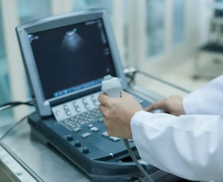
This examination helps investigate congenital heart problems before birth and evaluate the baby’s heart function. Fetal echocardiography focuses on the baby’s heart in greater detail than a standard pregnancy ultrasound. It is planned by the physician in pregnancies where it is considered necessary. Ionizing radiation is not used during the examination.
Based on the findings obtained, an appropriate plan for follow-up during pregnancy and postnatal evaluation can be created when necessary.
Normal Values in an Echocardiography Report
Normal values in an echocardiography report cover different measurements used to evaluate the structure of the heart and how it functions. The report may include data on the size of the heart chambers, wall thickness, contraction strength of the heart, valve structure, and blood flow. The ranges considered normal for these values may vary depending on the parameter measured, the person’s age, sex, body size, and the assessment method used.
Some of the measurements commonly seen in the report include:
- Ejection fraction: This shows how much blood the left ventricle pumps out with each contraction. In general, a range of approximately 50–70% is considered normal.
- Heart chamber measurements: The sizes of the left and right ventricles and atria are evaluated.
- Heart wall thickness: The heart muscle is examined for thickening.
- Valve assessment: The opening and closing movements of the valves are examined, along with possible narrowing or leakage.
- Doppler measurements: The direction and speed of blood flow within the heart are evaluated.
A single value in the report being outside the reference range is not sufficient on its own to make a diagnosis. The results are evaluated by the physician together with the other echocardiography findings and the person’s health status.
Frequently Asked Questions About Echocardiography
An echocardiogram (Echo) uses ultrasound waves to create images of the heart and evaluate its structure, valves, and function. An ECG records the electrical activity of the heart. Therefore, the two tests assess different aspects of heart health.
An echocardiogram provides information about the structure and function of the heart, but it does not directly show blockages in the coronary arteries. Depending on the clinical situation, additional tests may be required to evaluate coronary artery blockages.
A standard echocardiogram usually takes approximately 15–30 minutes. The duration may vary depending on the scope of the examination and the individual’s condition.
Fasting is generally not required before a standard echocardiogram, and you can usually eat and drink as normal. However, if a specific type of echocardiogram or another procedure is planned at the same time, the healthcare provider may give you specific preparation instructions.