

Color ultrasound is a specialized imaging technique used to analyze blood flow and direction within the vessels. With this method, potential blockages, narrowings, or flow disorders in the circulation can be easily detected.
Color ultrasound is especially preferred in pregnancy monitoring, evaluating cardiovascular diseases, and examining the structure of vessels that reach the organs. As it is a painless procedure and does not contain radiation, it provides safe and quick results. In this respect, it has become an indispensable part of the diagnostic process in many medical fields.
The key advantages of this method are as follows:
- Vascular mapping: The structures of vessels leading to organs can be examined in detail.
- Flow measurement: The direction and speed of blood flow can be clearly analyzed.
- Early diagnosis: Possible circulatory disorders can be quickly identified.
- Pregnancy safety: Since it does not involve radiation, it can be safely applied during pregnancy.
What is Color Ultrasound?
Color ultrasound is an advanced diagnostic method used to visualize blood flow in the vascular structures of the body. This procedure allows physicians to analyze the direction and speed of blood cells inside the vessels. Thanks to Color Doppler technology, not only the organ structures but also the vessels supplying these organs can be evaluated in detail. This enables the early detection of many vascular diseases.
During the imaging process, the device emits high-frequency sound waves. These waves reflect back after hitting body tissues, and the device converts these echoes into images. The direction and speed of blood flow are calculated based on changes in these sound waves. No pain is felt during the procedure, and it is typically completed within a few minutes.
So, how is the ultrasound performed?
- First, the patient is positioned appropriately based on the area to be examined.
- Then, gel is applied to the skin to ensure contact with the probe.
- The probe is directed toward the targeted vessels to capture images.
- Throughout the entire process, the patient remains in a comfortable position while the physician evaluates the images on screen.

Color ultrasound is used in various fields, from pregnancy to heart diseases. It is particularly effective in diagnosing conditions like vascular blockages, clot formations, or circulatory disorders. In this regard, it offers more comprehensive data compared to conventional ultrasound.
Color ultrasound is also frequently used during pregnancy. Evaluating fetal blood circulation is critically important, especially when monitoring placental and umbilical cord blood flow. This allows early detection of growth retardation, nutritional issues, or congenital heart anomalies. Experts use this imaging method periodically in high-risk pregnancies to closely monitor maternal and fetal health.
Why is Color Doppler Ultrasound Performed?
Color Doppler ultrasound is an imaging method that helps evaluate tissue structures as well as blood movement within the vessels.
Standard ultrasound and Doppler ultrasound serve different assessment purposes and should not be confused with each other. Standard ultrasound provides information about the shape and size of organs and their tissue characteristics. Doppler ultrasound helps examine the direction and characteristics of blood flow within the vessels. Accordingly, the examination may be planned particularly when blood supply in a specific area is thought to be reduced, increased, or irregular.
The main reasons for performing color Doppler ultrasound include:
- Assessing blood flow in arteries and veins
- Investigating suspected vascular narrowing, blockage, or blood clots
- Examining the distribution of blood vessels around organs, nodules, or masses
- Monitoring circulation after a treatment or surgical procedure
- Assessing blood flow in the placenta, umbilical cord, and fetus during pregnancy
Vascular examination may be necessary when findings such as swelling, pain, skin discoloration, or visibly prominent veins are present in the legs. It may also be used to support diagnostic assessment in different areas, including the neck, abdomen, kidneys, liver, thyroid, breasts, and testes.
Color Doppler ultrasound alone is not sufficient to establish a definitive diagnosis. The findings should be evaluated together with the physical examination, laboratory results, and other imaging methods when necessary. The area to be examined and the scope of the procedure are planned according to the existing symptoms and the medical requirements identified during the physician’s assessment.
When is Color Doppler Ultrasound Requested?
Color Doppler ultrasound may be requested when symptoms or previous assessments suggest a change related to blood circulation.
Unilateral swelling, pain, increased warmth, or skin discoloration in a leg may require an investigation to determine whether a blood clot is present in a vein. Leg pain that occurs while walking, coldness, numbness, or non-healing wounds may indicate the need to assess arterial circulation.
Other situations in which color Doppler ultrasound may be requested include:
- Suspected narrowing or blockage in the neck arteries
- Suspected varicose veins, venous insufficiency, or dysfunction of the venous valves
- Investigating suspected vascular dilation or an aneurysm
- Assessing the blood supply of an organ or tissue
- Examining the distribution of blood vessels around a mass, nodule, or lesion
- Performing follow-up after vascular surgery, stent placement, bypass surgery, or varicose vein treatment
- Assessing blood flow in the placenta, umbilical cord, and baby during pregnancy
The examination may be planned for the vessels in the neck, arms, legs, and abdomen, as well as for different areas such as the kidneys, liver, thyroid, breasts, and testes.
The need for color Doppler ultrasound is not evaluated solely on the basis of current symptoms. Physical examination findings, previous tests, existing medical conditions, and follow-up requirements are also taken into account. The area to be examined and the scope of imaging may vary depending on the medical need determined by the physician.
What Are the Prices of Color Ultrasound?
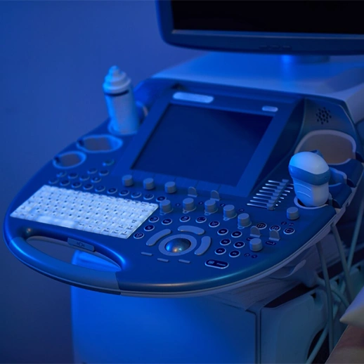
Color ultrasound is a diagnostic imaging method that enables a detailed analysis of the vascular system and blood flow. However, the pricing may vary depending on several factors. The equipment level of the healthcare institution, the experience of specialist physicians, and the quality of the technology used all influence the cost. Especially ultrasounds targeting leg circulation may require more detailed scanning, which can affect the pricing.
Main factors affecting the price:
- Whether the imaging is done at a private hospital or a clinic
- The scope of the area to be examined and the procedure duration
- The technological infrastructure of the device
- The field experience of the performing specialist
This imaging method is not limited to vascular assessments; it is also frequently used during pregnancy. In particular, examining intrauterine circulation in detail is vital to monitor conditions that may affect maternal and fetal health. These evaluations clarify whether the baby is receiving adequate oxygen and nutrients, reducing prenatal risks.
Moreover, some healthcare institutions offer Color ultrasound as a package service based on the body part to be scanned and the duration of the procedure. This provides flexibility in determining the cost. The most accurate information can always be obtained by directly contacting the relevant institution.
Following your doctor’s recommendation before imaging can provide advantages in terms of both time and cost. Especially for non-routine procedures, acting upon a doctor’s guidance will improve diagnostic accuracy.
Frequently Asked Questions
In pregnancy, Color ultrasound is usually applied from the 20th week onward. However, it can be done earlier or later depending on the doctor’s decision.
In Color ultrasound, blood flow within the vessels, its direction, and speed are evaluated. The circulatory system and vascular health are particularly assessed.
No, they are not the same. While detailed ultrasound evaluates the anatomical structure of organs, Color ultrasound analyzes blood flow and vessels.
Appointments for Color ultrasound are usually made through the radiology department. In some cases, specialists in gynecology or cardiovascular surgery may also provide referrals.
To get detailed information about Color ultrasound and plan your procedure, you can contact Denge Tıp.
Color Doppler ultrasound is an imaging examination that uses sound waves to visualize blood flow and vascular structures. It does not involve ionizing radiation during the examination. The procedure and evaluation process may vary depending on the area being examined and the clinical purpose of the study. Additional information can be obtained from the healthcare provider performing the examination.
The duration of a Color Doppler ultrasound depends on the anatomical region being evaluated, the scope of the examination, and the number of images required. Many examinations are completed within a relatively short period, although more comprehensive assessments may take longer.
Preparation requirements for a Color Doppler ultrasound vary depending on the area being examined. Fasting may be requested before evaluations of the abdomen or certain abdominal blood vessels. However, examinations of the thyroid, carotid arteries, breast, or leg veins often do not require fasting. Patients should follow the preparation instructions provided before their appointment.
After the examination, the acquired images are reviewed and a report is prepared. The time required to receive the results depends on the healthcare facility, the complexity of the examination, and the reporting process. In some facilities, results may be available on the same day, while in others, additional time may be required.