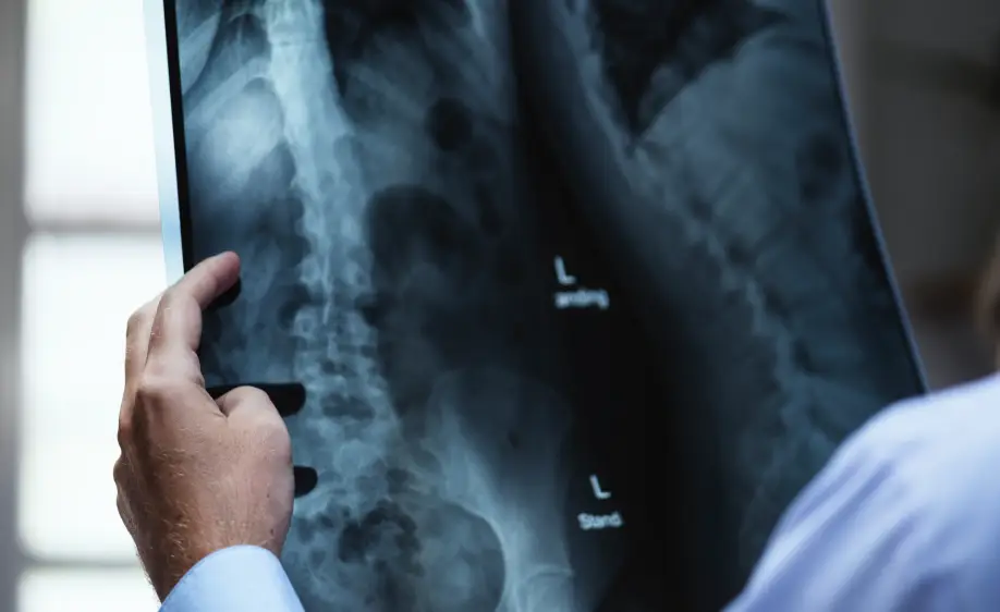
X-ray film is one of the most common tools used in the field of medical imaging. This film allows for a detailed examination of the internal structures of the body. It is especially preferred for assessing the condition of bones and organs. Through X-ray film, doctors can diagnose diseases early and determine the appropriate treatment methods.
X-ray film works on the principle of obtaining images using X-rays. As these rays pass through different parts of the body, they are absorbed differently depending on tissue densities, which appear as white, gray, and black tones on the film. It plays a crucial role in detecting fractures, tumors, or other abnormal conditions.
Conditions Diagnosable by X-Ray Film:
- Bone fractures and dislocations
- Joint inflammations and osteoarthritis
- Lung infections (such as pneumonia, tuberculosis, etc.)
- Lung cancer
- Bowel obstructions and stomach issues
- Heart enlargement
- Certain tumors

Foot X-Ray Film
Foot X-ray film is a common medical imaging method used to visualize the bones, joints, and soft tissues in the foot area. This method helps detect foot fractures, dislocations, bone deformities, and joint disorders. Foot X-rays are particularly useful for examining issues arising from sports injuries, falls, or chronic conditions. The images guide orthopedic specialists in making accurate diagnoses and planning treatments. Additionally, structural deformities in the bones of the foot and joint wear can be easily observed with this method.
X-ray films are not limited to the foot but also play a vital role in diagnosing conditions like scoliosis in the spine. Scoliosis refers to the abnormal curvature of the spine, and a full-spine X-ray is often required to assess this condition accurately. These images help determine the severity and progression of scoliosis.

Chest X-Ray Film
Chest X-ray film is a widely used medical imaging method to examine the organs and structures in the chest area. Chest X-rays are essential for diagnosing many respiratory diseases such as lung infections, lung cancer, pneumonia, tuberculosis, and bronchitis. It also helps identify conditions like heart enlargement and fluid accumulation in the lungs. Doctors often request a chest X-ray when patients experience respiratory problems.
Chest X-ray films can be taken while standing or lying down. The images provide valuable information about the size, shape, and tissue density of the lungs. This film allows doctors to create more effective treatment plans. Regular X-ray screenings are recommended, particularly for smokers, to detect lung cancer in its early stages.
Why is an X-Ray Film Taken?
X-ray films are taken to visualize bones and soft tissues in the body. These images, obtained using X-rays, are crucial in diagnosing pathological conditions such as fractures, dislocations, tumors, and infections. They are also commonly used in dental health, chest diseases, and orthopedic evaluations.
Since X-rays contain low radiation, they are a quick and reliable diagnostic method. X-rays are often preferred to quickly assess patients’ conditions in emergency medical interventions. Additionally, with the help of contrast agents, organ and vascular structures can be examined.
How Long Does It Take for X-Ray Films to Be Ready?
X-ray films are taken quickly, and in digital systems, the results can be viewed within minutes. However, in traditional film technology, this process can take 15-30 minutes, including film processing and development.
Thanks to digital X-ray technology, images can be examined quickly on a computer, and doctors can diagnose shortly after. However, interpreting and reporting the results by a radiologist may take some time, usually completed within a few hours.
Is MRI and X-Ray the Same Thing?
MRI (Magnetic Resonance Imaging) and X-ray are imaging methods based on different technologies. While X-rays visualize dense structures in the body (e.g., bones) using X-rays, MRI provides more detailed soft tissue images using radio waves and magnetic fields.
X-ray is fast and cost-effective. However, MRI offers higher resolution, especially for examining soft tissues such as the brain, spinal cord, muscles, and ligaments. MRI takes longer and does not involve radiation, while X-rays are completed in a shorter time but expose the patient to low doses of radiation.
Can Infections Be Detected on X-Ray?
Infections can be indirectly visible on X-ray. Particularly in lung infections, conditions like pneumonia can be detected using X-ray. Infected areas may show inflammation, fluid buildup, or abnormal tissue structures, which can be noticeable on an X-ray image. However, not every infection is clearly visible on X-ray.
Soft tissue infections or small infection foci may require more detailed imaging methods like MRI or ultrasound. Doctors combine X-ray results with other findings to make a diagnosis.


