Where is a chest X-ray taken is a frequently asked question in cases where structural changes in the chest area need to be examined radiologically. The procedure is usually performed in the radiology departments of hospitals or authorized imaging centers.
Where is a chest X-ray taken may vary depending on the purpose of the diagnostic process and the facility’s equipment. When necessary, the imaging procedure can be performed in the relevant departments upon a physician’s referral. The scan, carried out by qualified technicians, provides the data required for the physician’s accurate evaluation.
Where Is a PA Chest X-Ray Taken?
Among the most commonly used imaging methods to assess the overall condition of chest structures is the PA chest X-ray. This examination is performed in private imaging centers or in the radiology departments of hospitals.
During the procedure, the patient stands upright, keeping a certain distance from the device, and is asked to take a deep breath. This ensures that the heart, lungs, and chest cavity are clearly visualized. Due to its quick application and low radiation dose, this method is widely preferred in medicine.
In the preliminary evaluation of many respiratory system conditions, PA chest X-rays play an important role. Doctors examine the obtained images to analyze the structural integrity, air distribution, and vascular density of the lungs.
The technique used also helps determine whether the airways are open or if any obstructions exist. The size of the heart, the position of the diaphragm, and possible deformities of the chest wall can also be evaluated through this film.
The main situations in which this imaging procedure is requested are as follows:
- Used as an initial screening tool during examinations for lung cancer symptoms.
- Helps identify the cause of complaints such as cough, shortness of breath, chest pain, or fever.
- Performed during routine preoperative tests and for patients suspected of having an infection.
- May be taken when structural changes in the chest area need to be assessed after trauma.
PA chest X-rays also provide insight into general lung diseases. The images can reveal conditions such as lesions, fluid accumulation, or volume loss. However, advanced examinations may sometimes be required for a definitive diagnosis.
The specialist physician evaluates the results together with the clinical findings to determine whether any pathological condition is present. This approach forms the basis for an accurate and safe diagnostic process.
Where to Get a Chest X-Ray in Ankara
People who wish to undergo diagnostic examinations of the chest area often ask where to get a chest X-ray in Ankara. In the capital, this procedure can be performed in independent imaging centers, as well as in the radiology departments of public and private hospitals.
Before the scan, the patient is positioned correctly in front of the device. During this stage, the technician provides breathing and posture instructions to improve image clarity. This allows detailed visualization of the heart and lung areas.
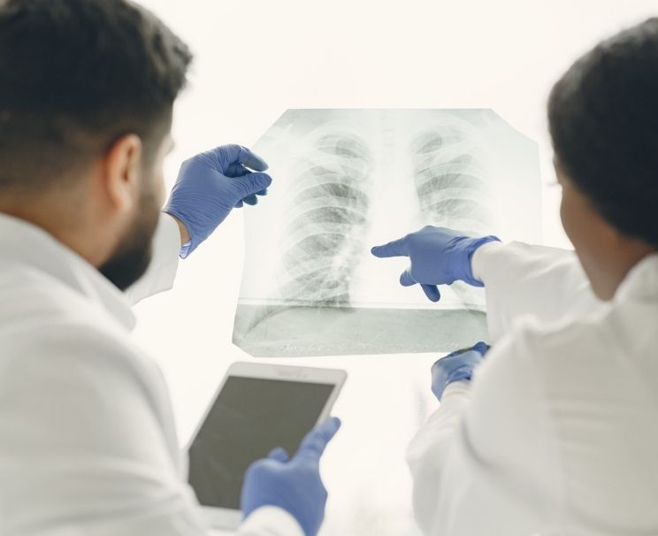
PA chest X-ray (PA CXR) is considered the first step in the diagnostic process. This film provides information about air distribution within the chest cavity, heart contours, and diaphragm position. Imaging also helps identify conditions such as infections, fluid accumulation, or volume loss.
The evaluation of the chest X-ray performed by a radiology specialist is based on interpreting the findings together with the clinical history. This approach supports physicians in making more accurate decisions during early diagnosis and treatment planning.
When a more comprehensive assessment is needed, computed tomography (CT) is preferred. This method provides sectional imaging of the chest tissues, allowing detailed analysis. It is also effective in clarifying cases involving suspected tumors or tissue deformities.
Key points to consider during the imaging process include:
- The patient must remain still in front of the device according to the technician’s instructions.
- Protective measures are taken to minimize radiation exposure.
- To enhance image quality, the patient should take a deep breath and remain motionless for a few seconds.
- Removing metal jewelry, watches, or accessories helps prevent reflections in the image.
The answer to how a chest X-ray is taken may vary depending on the patient’s age, physical condition, and the purpose of the scan. In some cases, the physician may request a CT scan of the lungs for more advanced analysis. This allows detailed examination of structural differences in the chest area and supports the definitive diagnostic process. To learn exactly what a chest X-ray is and in what situations it is requested, you can take a look at our article on Chest X-ray.
If you are looking for detailed information about where chest X-rays are taken, you can contact Denge Tıp for further assistance.