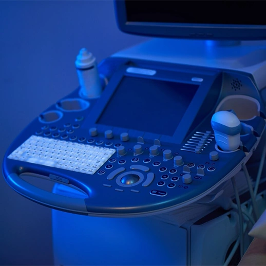
Ultrasonography is a safe examination technique that uses high-frequency sound waves to obtain detailed images of organs and tissues in the human body. Since it does not involve radiation, it is frequently preferred for pregnancy monitoring, abdominal organ examinations, vascular evaluations, and soft tissue analysis.
Ultrasonography provides real-time results during imaging. This allows physicians to make the necessary evaluations instantly during the examination. It can be used to examine many areas such as the abdominal region, thyroid, musculoskeletal system, vessels, and pelvic organs. In addition, Doppler ultrasonography can measure blood flow. With its non-invasive structure, this method preserves patient comfort while supporting the diagnostic process as a reliable tool.
Ultrasonography in Pregnancy
Ultrasonography is one of the most frequently used imaging methods to evaluate the health of the expectant mother and the development of the baby during pregnancy. Since it does not involve radiation and provides quick results, it is considered a safe option.
Throughout pregnancy, screenings performed at different weeks can assess the baby’s structural characteristics, organ development, and possible anomalies.
In answer to the question “How is ultrasound performed?”, a special gel is applied to the skin during the procedure, and sound waves are transmitted into the body with the help of an ultrasound probe. The reflected waves are converted into images by the device and examined by the physician. In this way, detailed information about the baby’s position, heartbeat, and development is obtained.

Examinations performed during pregnancy make it possible to:
- Detect many diseases at an early stage.
- Identify congenital anomalies.
- Examine the expectant mother’s pelvic organs, uterus, and ovarian structures.
- If necessary, evaluate the blood flow of the baby and placenta with color Doppler ultrasound.
Among the types of ultrasound used in pregnancy follow-up, the most common are 2D, 3D, and 4D ultrasounds. In addition, Doppler techniques provide information about the vascular structure and whether circulation is present. This method can also be used not only in pregnancy but also to examine gallbladder stones or polyps, as well as the liver, kidneys, and bile ducts.
Ultrasonography can also assist in diagnosing malignant formations. The main goal here is to monitor the health of the mother and baby during pregnancy. Regular ultrasound check-ups contribute to the early detection of possible risks, allowing the necessary medical precautions to be taken on time.
At Which Week Is Ultrasound Performed During Pregnancy?
During pregnancy, ultrasound is usually performed at specific weeks. The first screening is performed between the 6th and 8th weeks to confirm the presence and placement of the pregnancy.
The second important screening is done between the 11th and 14th weeks, and at this stage, the baby’s development and possible chromosomal risks are evaluated. The detailed ultrasound performed between the 18th and 22nd weeks is a critical period for examining organ structures.
Ultrasound is a safe imaging method that visualizes internal organs and tissues using high-frequency sound waves. During the procedure, a water-based gel is applied to improve contact between the probe and the skin, enhancing image quality. This gel helps obtain clearer images by improving the contact between the probe and the tissue.
During pregnancy follow-up ultrasound, examinations can be conducted to:
- Take measurements regarding the baby’s development.
- Evaluate the condition of the placenta and amniotic fluid.
- Monitor circulation with Doppler techniques that examine blood flow.
The examination referred to in medical literature as “detailed ultrasound” is especially performed around the 20th week. The information obtained in this way helps determine whether the pregnancy is progressing healthily. At the same time, it contributes to the early diagnosis of conditions that may affect maternal health.
Frequently Asked Questions
No, ultrasound does not involve radiation, and for this reason, it can be safely used in pregnancy and in examinations of different organs.
It is generally performed at least three times according to the trimesters, but it may be done more frequently if deemed necessary by the physician.
3D ultrasound provides static images, while 4D ultrasound offers moving, real-time images.
It evaluates the blood flow of the baby and placenta, providing detailed information about the circulatory system.
The area examined, the technology of the device used, and the type of test performed determine the cost.
Fasting may be necessary for abdominal ultrasounds; however, no special preparation is usually needed for pregnancy ultrasounds.
Gebelik sürecinizde güvenilir ve bilimsel yöntemlerle ultrasonografi hizmeti almak için Denge Tıp ile iletişime geçebilirsiniz.


