Chest X-ray is a fundamental diagnostic method that allows visualization of the structures within the chest cavity. This procedure helps physicians gather crucial information during the diagnostic process by evaluating the general appearance of the lung tissue, heart, and surrounding structures.
Chest X-ray is used to investigate possible infections, fluid accumulation, or structural abnormalities in individuals with respiratory complaints. During the imaging process, the patient usually stands upright and holds their breath briefly, preventing motion-related blurring. The obtained images guide physicians in assessing the overall health of the respiratory system.
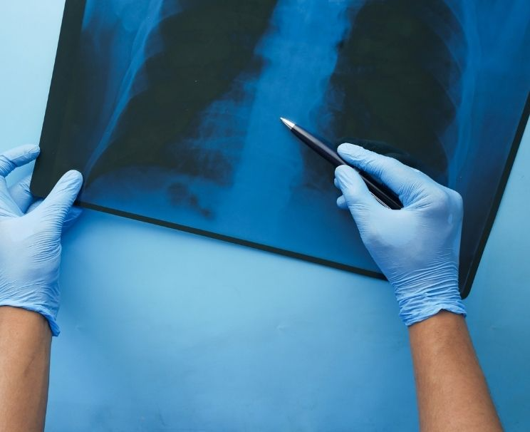
What is a Chest X-Ray?
A chest radiograph is a basic imaging technique used to examine the structural features of organs within the chest area. This examination provides general information about the lung tissue, heart shadow, and surrounding structures.
Physicians use this method to evaluate respiratory symptoms, monitor disease progression, or conduct preoperative assessments. Also known as a lung film, this is a reliable imaging technique commonly preferred in the initial stage of diagnosis.
The chest X-ray is produced when ionizing rays pass through tissues and are recorded by special detectors. In this way, conditions such as lesions, fluid buildup, or volume loss in the lung tissue can be detected. Images are usually taken in two directions (posteroanterior and lateral), allowing more accurate visualization of potential pathologies from different angles.
Some key features that make the thoracic X-ray valuable in the diagnostic process include:
- The procedure is very brief, and the patient is usually asked to hold their breath for a short time.
- It is painless and requires no incision or intervention.
- It simultaneously shows the general state of the lung tissue, heart size, and chest structure.
- The physician evaluates the obtained images alongside clinical findings to determine possible diseases.
Images acquired through radiography are interpreted by radiology specialists. These experts analyze the normal anatomical appearance of the lungs and any deviations from it. This process enables the initiation of an appropriate diagnostic plan and, if necessary, further tests.
Chest imaging, due to its rapid application and informative value, has become indispensable in modern medicine.
How is a Chest X-Ray Taken?
Chest imaging is one of the most frequently used diagnostic methods for examining respiratory system disorders. The procedure uses a low dose of ionizing radiation to make tissue densities inside the body visible.
The imaging process must follow specific technical steps to ensure accurate results. Proper positioning of the patient and breath control during the diagnostic procedure are crucial factors directly affecting image quality.
If you are considering having a chest X-ray, you can find detailed information about the process in our article Where to Get a Chest X-ray?
This examination is usually performed in a radiology unit using specialized equipment. The patient is placed in front of the machine, either standing or seated, and asked to hold their breath for a short moment. During this time, the chest area is exposed to radiation from a specific angle, and the reflected data are digitally recorded. The radiology technician ensures that the image is captured from the correct angles so that the organ boundaries appear clearly.
Key points to consider during thoracic imaging include:
- The patient should remove any metallic accessories before the procedure.
- Maintaining a fixed position prevents image blurring.
- A lead apron is used to avoid unnecessary radiation exposure to other parts of the body.
- For children and pregnant individuals, radiation dose is limited through special protective measures.
The posteroanterior (PA) chest X-ray evaluation process plays a crucial role in diagnosis. The specialist examines density differences in the image to identify possible infections, fluid buildup, or structural changes.
The standards to be followed during the procedure are not merely technical details but represent the quality criteria essential for accurate diagnosis. This imaging process requires careful planning and a skilled team to ensure precise results. To learn how the results are evaluated after a chest X-ray is taken, you can review our article, “Interpreting a Chest X-ray.”
Frequently Asked Questions
How should a healthy chest X-ray appear?
In a healthy image, lung fields appear uniformly dark without any increased density or opacity. The borders of the heart and diaphragm are distinct, and no pathological shadowing is seen within the lung tissue. Such an appearance is considered consistent with normal respiratory function.
What does whiteness on a chest X-ray indicate?
Whiteness in the image indicates that the tissues in that area are denser than normal. This may result from infection, fluid accumulation, a mass, or fibrotic changes. Clinical findings and additional examinations are evaluated for a definitive diagnosis.
Can pneumonia be seen on a chest X-ray?
Pneumonia generally appears as noticeable shading or opacity due to inflammation in the lung tissue. However, the image alone is not sufficient for diagnosis. The physician reaches a definitive conclusion by evaluating examination findings and laboratory results together.
For appointment requests and further information about chest X-ray services, you can contact Denge Tıp.