
Breast ultrasound is a medical and technological device that provides detailed imaging of breast tissues and structures. This method is used when clear results cannot be obtained with mammography. It is preferred for examining the nature of masses in cases where breast tissue is dense.
Breast ultrasound is a non-invasive procedure and is performed comfortably. A gel is applied to the area to be examined, and an ultrasound device is moved over the breast to obtain images. When used in conjunction with mammography, this method plays an important role in the evaluation and monitoring of breast health.
How is a Breast Ultrasound Performed?
A breast ultrasound is performed by a specialist radiologist or sonographer in the radiology department. This procedure involves the examination of breast tissue and the detection of possible abnormalities.
During ultrasonography, high-frequency sound waves are sent using an ultrasound device, and the returning waves are displayed on a monitor. The ultrasound image helps determine the structure and characteristics of masses.
The stages of the procedure are as follows:
The patient undresses from the waist up, exposing the area to be examined, and lies down on a stretcher.
A water-based gel is applied to the breast to ensure better contact with the ultrasound probe and clear images.
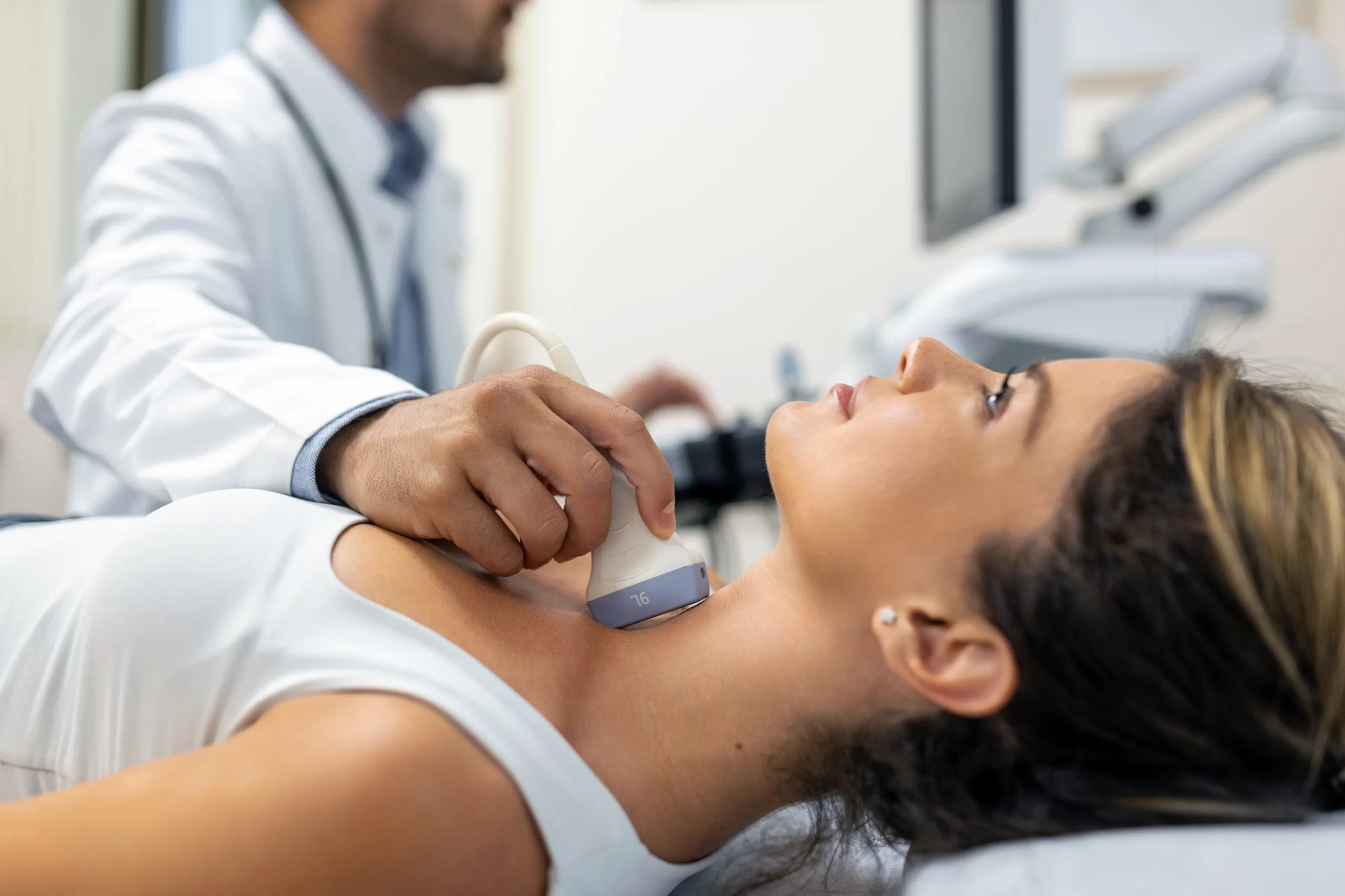
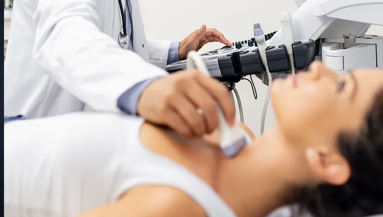
The radiologist or sonographer moves the ultrasound probe over the breast, sending sound waves. The probe detects the echoes from the tissues, and these signals are displayed on a screen.
The obtained ultrasound images are viewed on the screen and recorded. Areas deemed necessary can be examined in more detail.
The radiologist evaluates the images and determines if there are any abnormalities. These images are then documented in a report.
After the procedure is completed, the gel is cleaned from the patient’s skin, and the patient leaves the procedure room after getting dressed.

How Long Does a Breast Ultrasound Take?
The procedure typically takes between 15 and 30 minutes. However, this duration may vary depending on the patient’s condition and the extent of the area to be examined. This procedure is particularly preferred in young women, those with dense breast tissue, or situations where clear results cannot be obtained with mammography.
The question “Breast ultrasound or mammography?” is often asked. This question is important for determining which method is more appropriate for the patient.
Mammography is used for breast cancer screening, while ultrasonography is used to evaluate the nature of masses and identify structures such as cysts. Both methods can complement each other