Mammogram is considered one of the most important screening methods in terms of women’s health. This method is a control process recommended to be performed periodically after certain ages. The aim is to detect possible risks at an early stage and ensure that treatment is started quickly.
Mammogram is performed at intervals determined under the guidance of specialist physicians, and the results obtained in routine check-ups are compared to detect possible changes. In this way, risk factors are not overlooked, and women’s quality of life is preserved. The advantage of early diagnosis increases the success of treatment and supports an easier recovery process. With the rise of public awareness, the importance of having this screening regularly is being emphasized more and more every day.
- Based on risk factors, mammogram is recommended for the following individuals:
- Women with a family history of breast cancer
- Individuals referred by their doctor due to previous health problems
- Women undergoing hormone therapy or those with dense breast tissue
- Individuals recommended to be included in a regular screening program after the age of 40
What is Mammogram?
Early diagnosis plays a highly significant role in women’s health. Breast X-rays help detect changes occurring in breast tissue. Through this method, small lumps or calcifications can be identified, creating the possibility of treatment at an early stage. Today, thanks to technological advances, more precise results can be obtained, and specialist doctors can carry out evaluations more reliably.
In the medical field, breast radiography is widely used; it reveals the internal structure of breast tissue with low-dose radiation. Regular check-ups make it easier to detect potential risks at an early stage. This method plays an important role not only in detecting suspicious findings but also in routine screenings.
Breast screening performed for early diagnosis increases the success rate in the fight against the disease, contributing to a shorter and more effective treatment process. Depending on the size of the lumps or the density of the tissue, specialists may recommend different methods.
The techniques used in this process offer various advantages:
- With high-resolution imaging devices, even small changes can be detected.
- The evaluations of radiology specialists are a critical step in reaching an accurate diagnosis.
- Regular check-ups provide personalized health monitoring.
- When tissues with different densities need to be examined, additional methods may be used to support the analysis.
This method is not only diagnostic but also an important part of preventive medicine. Routine check-ups contribute to the protection of women’s health. Therefore, without ignoring the power of early diagnosis, it is crucial to continue screenings as recommended by specialists.
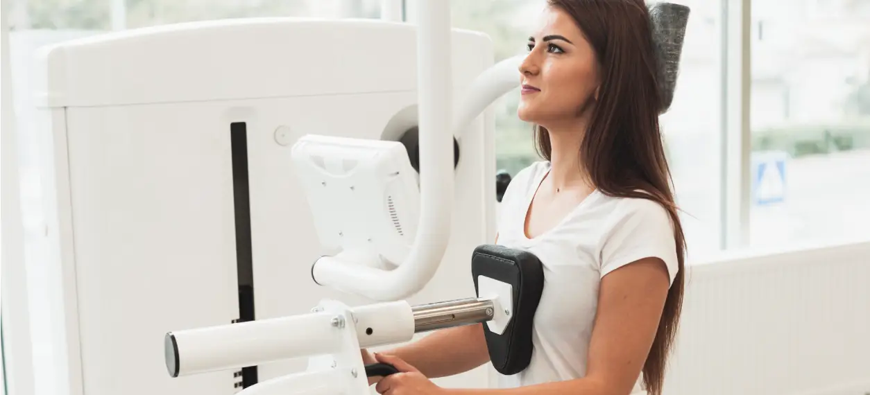
How is Mammogram Performed?
One of the methods used to examine breast tissue is digital mammogram, which is among the most preferred techniques today. This application allows images to be stored digitally and examined in detail. It is considered one of the most important steps in the timely detection of breast cancer and is recommended to be performed at certain intervals after a certain age.
In general, these screenings start at the age of 40, but for women in risk groups, they may be recommended much earlier. The imaging process is performed with a low radiation dose, which is a reassuring feature for patients in terms of safety. Thanks to the technical capacity of the mammogram device used, clearer results can be obtained.
To ensure the most accurate results in the breast area, the period when the breasts are least sensitive is chosen. Thus, possible pain or discomfort is minimized. These examinations are carried out not only to evaluate suspicious findings but also for screening purposes. In this way, risky conditions can be detected before symptoms appear.
The process works as follows:
-
- During mammogram, the breast tissue is compressed and stabilized between special plates.
- Short-term pressure ensures the clearest possible images.
- Screenings performed for the early detection of breast cancer may contribute to the timing of treatment.
- Removing jewelry and wearing comfortable clothing before the procedure makes it easier to perform.
For more detailed information and to find out whether these check-ups are suitable for you, you can contact Denge Tıp.