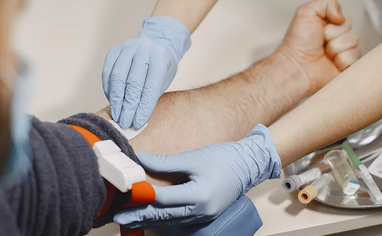
HSG film imaging is a radiological imaging technique used to evaluate the structural condition of the uterine cavity and fallopian tubes. Specialists inject contrast material into the uterus to clearly visualize the uterine cavity and the patency of the tubes via X-ray.
HSG film imaging is usually performed within the first week following menstruation, and a gynecological examination is carried out beforehand. Some women may feel discomfort similar to menstrual cramps when the contrast material is administered. Patients can typically resume daily activities within a few hours after the imaging. Some patients may experience slight bleeding or spotting afterward; doctors generally consider this normal.
How Is HSG Film Imaging Performed?
HSG film imaging, medically known as hysterosalpingography, is a special imaging technique used by doctors to assess the structure of the uterus and fallopian tubes. It is especially used in women planning pregnancy but facing difficulties, as it allows specialists to examine the shape of the uterine cavity and tube patency in detail.
A contrast agent is used to clearly examine the structure of the uterus and fallopian tubes. This application is generally performed under X-ray guidance by a physician.
To ensure a safe and comfortable experience, some preparations are essential before the imaging. The best time for an HSG film is a few days after menstruation ends. Therefore, patients should consider their menstrual cycle when scheduling an appointment to get a clear answer to the question of when the uterine film should be taken. This helps prevent the possibility of pregnancy and minimizes the risk of infection.
The imaging is carried out in the following steps:
Mild vaginal discharge or spotting may occur afterward. Patients should be informed and advised to rest after the imaging. If needed, doctors may prescribe pain relievers or antibiotics.
In some cases, the contrast material may not spread completely within the uterus. This may indicate blocked tubes or congenital anomalies in uterine structure. Accurate interpretation of HSG results is crucial for planning further investigations or treatments.
Where Is HSG Film Imaging Performed?
HSG film imaging is one of the detailed imaging images related to female reproductive health. It is performed to examine the structural condition of the uterus and fallopian tubes and is conducted in radiology clinics equipped with special devices.
To ensure safety and accurate results, it is recommended that the imaging be done in centers with experienced radiologists. During the imaging, contrast material is injected to make the uterine structure and tubal patency visible. Therefore, device quality and practitioner experience are very important.
For uterine film imaging, centers that meet hygiene standards and offer advanced technical infrastructure should be preferred. Hospital radiology departments, university hospitals, and imaging units within obstetrics clinics are capable of performing this imaging.

The imaging is usually planned right after the menstrual period. At this time, the uterine cavity is empty, and the possibility of pregnancy is eliminated. During the imaging, the patient lies in a gynecological position, contrast material is administered via a thin catheter through the cervix, and sequential images are obtained using an X-ray device.
There are certain things patients should be mindful of before and after the imaging. Prior infections or chronic diseases must be reported to the doctor. Informed consent is obtained after a pre-imaging consultation.
Precautions include maintaining hygiene, using pain relievers for mild discomfort, and allocating a few days for physical rest. On the day of the imaging, comfortable clothing should be worn, and patients should inform their doctors about any regularly used medications.
How Is a Uterine Film Taken?
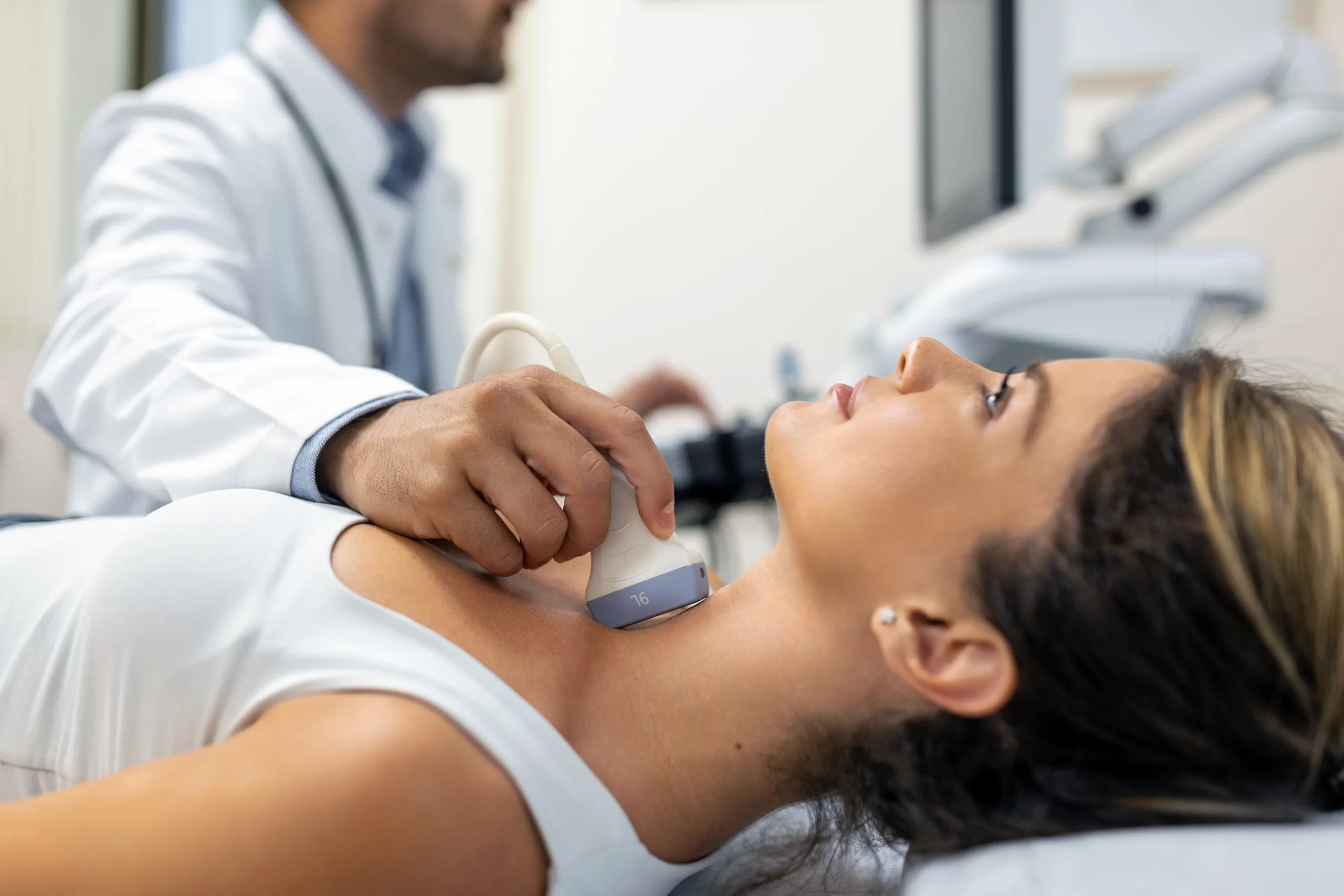
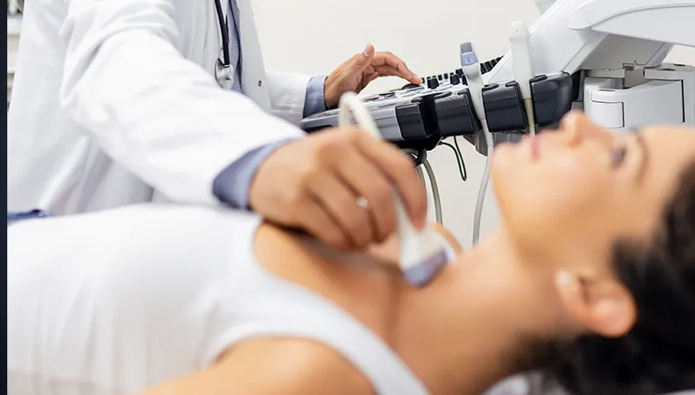
In this imaging, the patient is positioned for a gynecological exam under sterile conditions. A speculum is inserted to expose the cervix. A thin catheter is then used to deliver contrast material into the uterus. The distribution of contrast within the uterine cavity and tubes is monitored in real-time with an X-ray device. This allows a clear evaluation of the uterine structure and tubal patency.
Is There an Allergy Risk in HSG Film Imaging?
In rare cases, patients may develop reactions to the contrast agent used during HSG film imaging. This risk should be considered, especially in individuals with a history of allergies. Before the imaging, any prior reactions to medications or contrast agents must be evaluated.
If an allergic reaction occurs, the medical facility must be able to intervene promptly. In such cases, treatment should begin immediately to ensure patient safety. Therefore, the center chosen for HSG film imaging should not only have technical adequacy but also the necessary equipment to ensure patient safety.
Frequently Asked Questions
Mild cramp-like pain may be felt during the HSG imaging, but it is generally brief and tolerable. Some patients may feel discomfort similar to menstrual cramps, though most tolerate the imaging well. Patients with low pain tolerance can take a pain reliever before the imaging upon their doctor’s advice. Mild abdominal pain or vaginal discharge may occur afterward, but this is temporary.
HSG işleminden sonra enfeksiyon riskine karşı genellikle 2 ila 3 gün ilişki önerilmez. Doktorun özel bir önerisi yoksa, vücut kendini toparlayana kadar cinsel ilişkiye bir süre ara verilmesi tavsiye edilir. Vajinal akıntı ya da hafif kanama bitene kadar beklenmesi enfeksiyon riskini azaltmak açısından faydalıdır. İyileşme sürecini desteklemek için hijyene dikkat edilmesi ve belirtiler gözlenmesi önemlidir.
An HSG imaging typically takes 10 to 15 minutes. Preparation and positioning may take additional time, but the actual imaging is brief. The total time spent at the clinic may be around 30 minutes. A short observation period may follow the imaging, depending on the patient’s condition.
You can contact Denge Tıp to get detailed information about HSG imaging.