

Bone scan refers to imaging and measurement methods used to evaluate structural changes in bone tissue, mineral density, or abnormalities related to bone metabolism. Bone density measurement is mainly used to assess osteoporosis and fracture risk. Bone scintigraphy helps examine the bones in more detail in cases where fracture, infection, tumor, or metastasis is suspected.
Bone scan is an important examination for detecting bone diseases at an early stage and creating an appropriate treatment plan. Bone density measurement allows bone loss to be identified and fracture risk to be assessed, especially in people at risk of osteoporosis. In patients with a history of cancer or suspected metastasis, this examination is also one of the important methods used to investigate whether the disease has spread to bone tissue.
Bone density measurement plays a role in diagnosing the following diseases:
- Osteoporosis (bone loss): Detects decreases in bone density and assesses fracture risk.
- Osteopenia: Identifies mild decreases in bone density that precede osteoporosis.
- Bone metastasis: Determines if cancer has spread to the bones.
- Bone tumors: Evaluates the presence or spread of tumors in the bones.
- Paget’s disease: Detects abnormal bone growth and deformities.
- Bone infections (osteomyelitis): Diagnoses bone inflammation.
- Fractures: Helps identify small, non-visible stress fractures.
How is a Bone Scan Performed?
A bone test is performed to assess the density and health of bones. It is commonly used to detect decreases in bone density, such as in osteoporosis. The most widely used method is the DEXA (Dual-Energy X-ray Absorptiometry) scan.
The DEXA scan is typically performed on the spine, hip, and wrist, which are the areas most prone to fractures. This method provides a detailed examination of the body’s bone structure, allowing early detection of bone loss.
During the test, the patient lies on a special table, and images are taken using low-dose radiation. These images measure bone mineral density and provide information about bone health. The test takes about 10-30 minutes and is painless.
This method helps detect bone loss early and assess fracture risk. The results are analyzed by doctors to guide the diagnosis and treatment of conditions such as osteoporosis.

In some cases, more detailed bone evaluation is required. In these situations, nuclear medicine techniques are used. In nuclear medicine, a radioactive substance is used to obtain detailed images of the bones. This method is used to detect more serious conditions, such as bone tumors, metastases, or bone infections.
Based on the bone test results, appropriate treatment and preventive measures are planned for the patient. A bone scan is a recommended health check, especially for older adults or those at risk.
| Stage | Description |
| Preparation Before the Procedure | Before a bone scan, the patient’s general health condition, medications used, and possible pregnancy status are evaluated. If the procedure is performed as bone scintigraphy, the patient may be asked to remove metal accessories. When necessary, the patient is given specific information before the procedure. |
| Radioactive Substance Administration | During bone scintigraphy, a low-dose radioactive substance is usually administered intravenously. This substance is taken up by bone tissue and helps abnormalities in the bones to be evaluated more clearly during imaging. |
| Imaging Process | A certain period of time is allowed for the radioactive substance to spread through the bone tissue. Then the imaging stage begins. The patient is positioned appropriately in the imaging device, and the areas of uptake in the bones are examined. The images obtained are evaluated by the physician and interpreted in terms of bone diseases other than osteoporosis, as well as suspected tumors or metastasis. |
Why is a Bone Scan Done?
The most common use of a bone scan is to detect conditions that lead to decreased bone density, such as osteoporosis. It is a critical method for evaluating fracture risk, especially in postmenopausal women and those at risk of bone loss. If bone density decreases are detected early, appropriate treatment and lifestyle changes can help protect bone health.
A bone test is also performed to detect cancer metastases in the bones, evaluate bone infections (osteomyelitis), and examine bone tumors. In these cases, a more advanced test, known as bone scintigraphy, may be used.
Bone scintigraphy uses a radioactive substance to provide detailed images of the bones. This method helps detect abnormalities in the bones early, speeding up the treatment process.
Bone density testing is also important in diagnosing conditions like Paget’s disease, which causes structural abnormalities in the bones. Nuclear medicine techniques such as scintigraphy and other bone scans help detect such conditions early, guiding patients to the appropriate treatment.

In Which Department is a Bone Scan Performed?
The department where a bone scan is performed may vary depending on the method used and the health problem being investigated. In general, bone density measurement is performed in the radiology department. This method evaluates bone mineral density and provides information especially about osteoporosis, osteopenia, or fracture risk. Bone density measurement may be requested by a physician for people in the postmenopausal period, older adults, those using corticosteroids for a long time, people with a previous fracture history, or individuals at risk of bone loss. Therefore, the procedure is not limited only to the imaging stage. It should be evaluated together with the patient’s general health condition and risk factors.
Bone scintigraphy, on the other hand, is an examination performed in the nuclear medicine department. It may be preferred to evaluate suspected bone metastasis, bone tumors, infection, unexplained bone pain, or certain fractures. The first medical specialty to consult before the procedure may vary depending on the patient’s complaint. Orthopedics, physical therapy and rehabilitation, oncology, endocrinology, or internal medicine physicians may refer the patient to the radiology or nuclear medicine unit when necessary. The department where a bone scan is performed is determined according to the type of test requested and the clinical evaluation.
How Are Bone Scan Results Interpreted?
Bone scan results are interpreted differently depending on the type of procedure performed. In bone density measurement, the main purpose is to evaluate bone mineral density and determine the person’s risk status in terms of osteoporosis or osteopenia. The values obtained through this examination are assessed together with the patient’s age, sex, menopausal status, history of previous fractures, medications used, and accompanying diseases. Low bone density in the results may indicate an increased fracture risk. However, the treatment decision is not made based only on the measurement result. The physician evaluates the patient’s overall risk profile and determines whether follow-up, lifestyle changes, or medication treatment is needed.
In bone scintigraphy results, the areas where the radioactive substance is taken up in bone tissue are examined. Increased uptake in some areas may require further investigation in terms of fracture, infection, tumor, metastasis, or different bone diseases. However, not every area of uptake seen on scintigraphy definitely means cancer or metastasis. These findings are evaluated together with the patient’s complaints, physical examination results, blood tests, and, if necessary, additional imaging methods such as MRI, CT, or X-ray. Therefore, bone scan results must always be interpreted by the relevant physician.
Is a Bone Scan Harmful?
A bone scan is an examination that may involve low-dose radiation depending on the method used. In bone density measurement, the amount of radiation is generally very low, and the procedure is completed in a shorter time. When deemed necessary by the physician, this method is considered safe for evaluating osteoporosis and fracture risk. In bone scintigraphy, a low-dose radioactive substance is administered intravenously. This substance is taken up by bone tissue and helps imaging to be performed. It is also eliminated from the body within a certain period of time. After the procedure, the patient should drink plenty of fluids or follow certain safety recommendations.
As with all procedures involving radiation, the patient’s general health condition should be evaluated before a bone scan. If there is a possibility of pregnancy, pregnancy, or breastfeeding, this information must be shared with the physician and healthcare team. During pregnancy, imaging procedures involving radiation may be postponed unless they are necessary, or different methods may be preferred. During breastfeeding, the physician decides whether short-term precautions are needed depending on the method used. Although bone scanning is generally a safe procedure, an individual assessment is necessary in terms of pregnancy, breastfeeding, and special health conditions.
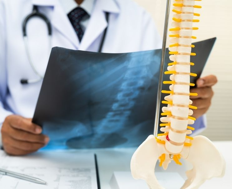
Frequently Asked Questions About Bone Scans
This procedure is generally not painful. During bone scintigraphy, there may only be a slight needle sensation when the substance is administered intravenously.
Bone density measurement is usually completed within a short time. In bone scintigraphy, the process may take longer because time is needed for the radioactive substance to spread through the bone tissue.
Fasting is generally not required for this procedure. However, depending on the method to be used and the patient’s condition, the physician or relevant unit may request special preparation before the procedure.
There may be low-dose radiation depending on the method used for the bone scan. The amount of radiation is very low in bone density measurement, while a low-dose radioactive substance is used in bone scintigraphy.
Bone scintigraphy and bone density measurement are not the same procedure. Bone density measurement mainly evaluates osteoporosis and fracture risk. Bone scintigraphy is used to investigate metastasis, tumors, infection, or different bone diseases.